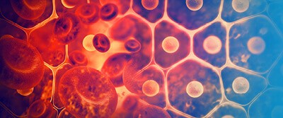
Peering Inside Living Cells

By Nancy H. Pitts
Think of a living cell as a terrarium: a self-contained, highly efficient environment that sustains life through complex processes. It’s fun to look inside. If you have tried to maintain and study a terrarium, you have probably learned that it’s very fragile. Simple exterior forces on its self-contained environment (opening it, temperature variations, too much or too little light or water, etc.) can damage the terrarium so that the plants inside die. Similarly, peering inside a living cell used to introduce exterior forces that damaged or killed the cell, until two engineers developed the “Ultrasound Bioprobe.”
Complex, invasive, destructive forces traditionally have been used to examine living cells. Staining, fixing, slicing, dicing, etc., either damaged or killed the delicate cell. A dead or damaged cell is not like a living cell. Think about that unsuccessful terrarium: it’s certainly not an example of thriving, on-going life.
Traditional methodologies for studying living cells had other limitations. Fluorescent microscopes offered poor resolution and distortion; electron microscopes were too destructive; light and wave imaging couldn’t view anything that small; and scanning probe microscopes couldn’t see inside.
Imaging a Living Cell Without Killing It
Gajendra Shekhawat and Vinayak P. Dravid, professors of materials science and engineering at Northwestern University, recently developed a method for looking inside, or ‘imaging’, living cells without damaging them. Their ultrasound bioprobe uses ultrasound imaging deep inside the living cell combined with atomic force microscopy for high-sensitivity, high-contrast resolution (wide range of data) images. The result: great pictures of the inside of a living cell.
What makes this exciting to scientists is that they can now examine a living cell in nanometer scale resolution. There are 25 million nanometers in one inch; there are 10 million nanometers in a centimeter.
Here is one result: The ultrasound bioprobe is helping scientists understand the tiny mechanical failures of living lung endothelial cells while they’re happening, further advancing the treatment of critically ill patients who develop a serious and deadly lung problem. This can help scientists measure the effectiveness of certain treatments and medications. Understanding living cell component failures and measuring the effect of treatment agents is a sure road to achieving success. Not bad, just for peeking inside.
Discussion Questions
- How do you become a materials science engineer?
- What are some other important outcomes of being able to study living cells?
Vocabulary
- Terrarium
- Ultrasound
- Microscopy
- Endothelial
