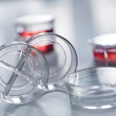
Great Fluorescence Microscopy Requires the Right Consumable
From the status of your cells to the settings of your microscope, many factors can influence the quality of your images in fluorescence microscopy. With so many factors in play, it’s important to choose the right vessel for your application to start your microscopy on the right foot. The vessel should have the correct bottom thickness, a planar surface, low autofluorescence, and the right surface chemistry, and it should also prevent cross talk and bleaching.
Bottom Thickness
Microscope objectives are typically calibrated to accommodate a standard cover glass thickness of 175µm. For microscopic applications at higher magnifications, a thicker vessel bottom can make it difficult to adjust the focus and significantly reduce resolution. This effect can be exacerbated with oil or water immersion objectives. Generally, choose a vessel with a bottom thickness of 175µm (#1.5 coverglass) to improve your image quality.
Autofluorescence
Autofluorescence refers to non-specific fluorescence that can occur with proteins, buffers, fixatives, and microplate or slide surfaces, so choose the proper plastic or glass vessel to minimize autofluorescence. For example, polyolefins and glass have low levels of autofluorescence and are ideal for high content imaging. A microplate with black wells can also help to quench any background autofluorescence.
Cross Talk and Bleaching
Well-to-well cross talk occurs when stray light reaches a different well during microscopy. Cross talk can cause bleaching or over illumination of samples in nearby wells. To counteract this, black well sides can block out light and guarantee that each cell produces its maximum signal strength at the beginning of the screening.
Surface Chemistry
Whether your vessel surface is tissue culture treated, has Advanced TC treatment, or is coated with protein, you want adherent cells to attach. The surface chemistry of untreated glass may not promote the attachment of your cells, but surface-treated plastic or glass will enhance the cell binding and improve their morphology. When performing cultures where attachment is not preferred, a vessel with a glass-bottom surface or one made from cell-repellent polystyrene may be ideal.
Planarity
A flat, planar surface provides quality images when refocusing is not an option. Planarity is important for clear, in-focus images, especially with high-speed and high-resolution microscopy.
Greiner Bio-One CELLview Dishes, Slides, and Plates can help you overcome these concerns. Contact your Fisher Scientific sales representative to try them in your application today.

Content provided by:


