Histology Workflow Products
Histology, a key component of anatomic pathology (AP), is key to tissue-based diagnoses and successful patient treatment.
Once an essentially manual process, automation is being introduced to the histology lab. Additionally, the histologist may now perform molecular diagnostics on tissue samples, and digital microscopy makes it easier and faster to share critical information with other clinicians.
Histology Workflow
A typical histology workflow consists of:
- Specimen Collection: tissue specimens delivered from the operating room or physician office are examined, dissected, and placed into labeled cassettes
- Tissue Processing: the cassettes are placed into a processor that dehydrates, clears, and infiltrates the tissue (process takes 10 to 12 hours)
- Tissue Preparation: the tissue is encased in paraffin or other media, sliced using a microtome, placed on microscope slides, stained, and coverslipped
Multiple slides from each sample are assembled as a “case” and forwarded to a pathologist for microscopic examination and evaluation.
Specimen Collection
During grossing, the pathologist measures, describes, and dissects the specimen and places pieces of the tissue into labeled plastic cassettes (blocks). Because the tissue may have been placed in formaldehyde or other tissue preservatives, working under a hood or enclosure is recommended.
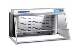
Grossing Station
Protect users from formaldehyde and other chemical exposure during grossing.
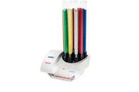
Cassette Label Printer
Manual or automated cassette labeling is key to proper specimen identification and tracking.
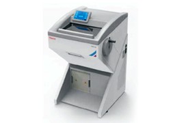
Cryostat
Process “frozen sections” quickly with cryotomes that combine a microtome-like cutting mechanism with a freezer unit.
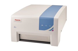
Storage Systems
Safely store and save time sorting, archiving, and retrieving your slides and tissue blocks.
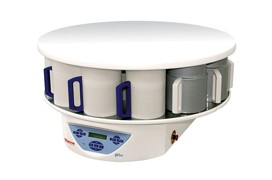
Tissue Processors
Automate the overnight process of subjecting tissue to a series of alcohol solutions, xylene clearing agents, and paraffin.
Tissue Preparation
Tissue preparation includes embedding, sectioning, staining, and coverslipping. The specimen is embedded in paraffin, the blocks are sliced using microtomes, and the tissue sections are placed onto glass microscopic slides. The slides are stained and then coverslipped to create a permanent preparation.
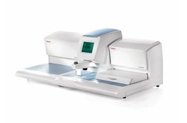
Tissue Embedding Station
Embedding stations facilitate the embedding process with cooling surfaces that rapidly harden paraffin.
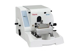
Microtome
Section tissues with manual, rotary, sliding, or automatic microtomes with reusable or disposable blades.
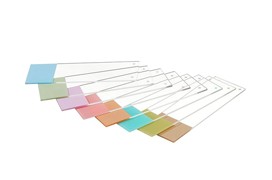
Slides and Slide Labeling
The right type of slide and accurate, reliable labeling are key to successful specimen mounting and identification.
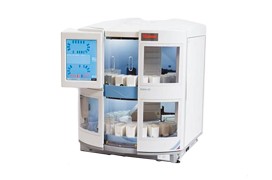
Stains and Reagents
Find standard Hematoxylin and Eosin (H&E) and other tissue and cell stains to help identify cellular components and features.
- Thermo Scientific Gemini AS Stainer
- Thermo Scientific™ Richard-Allan Scientific™ Signature Series Clear-Rite™ 3
- Thermo Scientific™ Shandon™ Instant Eosin
- Thermo Scientific Richard-Allan Scientific Signature Series Eosin-Y 7111
- Thermo Scientific Richard-Allan Scientific Signature Series Bluing Reagent
- Thermo Scientific Richard-Allan Scientific Signature Series Hematoxylin 7211
- Thermo Scientific Richard-Allan Scientific Signature Series Cyto-Stain
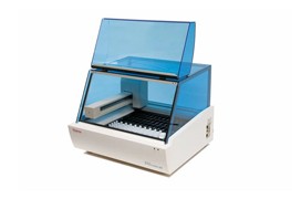
Immunohistochemistry
Choose reagents and antibodies to help identify cellular antigens and markers of specific tissue and tumor types.
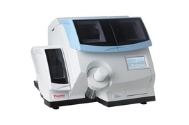
Coverslips
Coverslips and mounting media make your tissue slides a part of the patient’s permanent record. Automated coverslippers help to eliminate tedious and labor-intensive steps.
