First True-Color Super-Resolution Microscopy Technique Developed
By Brianne McCurley
To say there have been many advancements in the field of microscopy since the development of the first compound microscope by Zaccharias Jansen in the late 16th century is an understatement. Jansen and his father, Dutch spectacle makers, built the first microscope by using three draw tubes with lenses inserted into the ends of the flanking tubes. They discovered a much larger image than expected; much larger than simple magnifying glass provided. The very first compound microscopes only magnified images between 6x – 9x. Microscopes of today can magnify images to the nanometer.
The SR-STORM, or spectrally resolved stochastic optical reconstruction microscopy, was developed by Ke Xu, a faculty scientist in University of California Berkeley Life Sciences Division and an assistant professor at UC Berkeley’s Department of Chemistry. This new type of imaging combines single-molecule spectral measurement with super-resolution microscopy. It provides full spectral and spatial information for each molecule by coloring each molecule according to its spectral position. Xu was able to build on earlier super-resolution techniques developed by Harvard professor Xiaowei Zhuang, who invented STORM, a super-resolution microscopy method based on single-molecule imaging and photoswitching.
According to Science Daily, the SR-STORM research was reported in the journal Nature Methods in a paper titled, “Ultrahigh-throughput single-molecule spectroscopy and spectrally resolved super-resolution microscopy,” with co-authors Zhengyang Zhang, Samuel Kenny, Margaret Hauser and Wan Li, all of UC Berkeley.

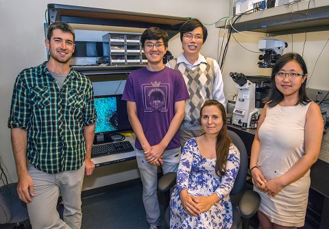
Ke Xu (center) and colleagues have pioneered a new super-resolutionmethodology to image single molecules with unprecedented spectral and spatial resolution (Roy Kaltschmidt/Berkeley Lab)
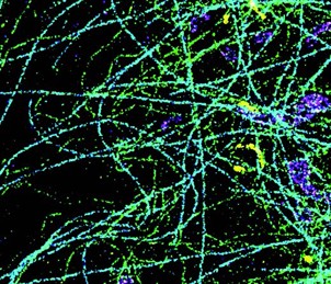
According to Xu, “We measure both the position and spectrum of each individual molecule, plotting its superresolved spatial position in two dimensions and coloring each molecule according to its spectral position, so in that sense, it’s true-color super-resolution microscopy, which is the first of its kind. This is a new type of imaging, combining single-molecule spectral measurement with super-resolution microscopy.”

Methodology: The Process Used By The Researchers
They constructed a dual-objective system with two microscope lenses facing each other.
Xu and colleagues viewed the front and back of the sample at the same time and achieved unprecedented optical resolution (of approximately 10 nanometers) of a cell.
Next, they dyed the sample with 14 different dyes in a narrow emission window, excited and photoswitched the molecules with one laser. (While the spectra of the 14 dyes are heavily overlapping since they’re close in emission, they found that the spectra of the individual molecules were surprisingly different and thus readily identifiable).
Then, they used four dyes to label four different subcellular structures, such as mitochondria and microtubules, and were able to easily distinguish molecules of different dyes based on their spectral mean alone, and each subcellular structure was a distinct color.
Researchers looked at the interactions between four biological components inside a cell in 3D and at very high resolution (about 10 nanometers). Xu stated that “the applications are mostly in fundamental research and cell biology at this point, but hopefully it will lead to medical applications.”
The SR-STORM allows one to view the cytoskeleton of a cell. “Using microscopy techniques, like SR-STORM, which include staining, researchers have been able to demonstrate the myriad forms which the cytoskeleton can take, and much has been learned about the architecture of the cytoskeleton.” These advances may someday help researchers and doctors understand more about diseases, such as Alzheimer’s, which is believed caused by the degradation of the cytoskeleton inside neurons.
Extension Questions:
Define these three types of microscopes and for what applications they are used.
- Optical
- Electron
- Scanning probe